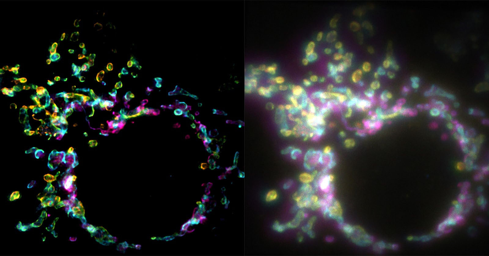
Super-Resolution Imaging with SPIFFI
A breakthrough in fluorescence microscopy offers scientists an unprecedented glimpse into cellular structures. The new technique, known as SPIFFI (spatial polarization-induced fluorescence fluctuation imaging), allows for super-resolution imaging from a single exposure, solving challenges associated with traditional methods.
Key Features of SPIFFI:
- Single Exposure Imaging: Unlike traditional super-resolution microscopy, which relies on capturing hundreds or thousands of images, SPIFFI achieves high resolution from just one image.
- Polarization Advantage: The technique leverages the polarized light emitted by fluorescent molecules, splitting this light into four channels to reveal hidden structural details.
- Resolution Improvement: SPIFFI can enhance image resolution by a factor of two, reaching resolutions of about 160-170 nanometers, with potential for even finer details down to 80 nanometers when integrated with existing methods.
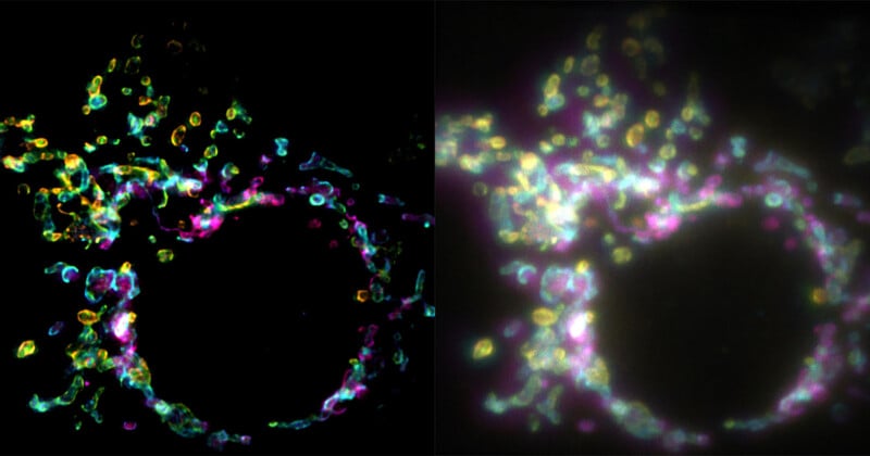
Mitochondrial outer membrane imaged with SPIFFI (L) vs widefield microscopy (R).
Significance:
- SPIFFI enables the observation of fast-moving cellular processes, making it possible to create super-resolution videos of live cells.
- Its optical hardware can be integrated into current fluorescence microscopes, enhancing practicality and accessibility in laboratories.
This innovative technique, detailed in a recent Nature publication by researchers Wei Guo, Lely Feletti, and Aleksandra Radenovic, represents a significant advancement in cellular imaging technology.
For more detailed insights, you can check out the full research paper.
Image Credits: EPFL