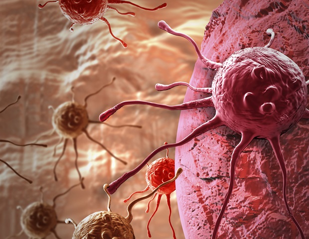
Recent research from the University of Warwick highlights significant concerns regarding the optimism surrounding artificial intelligence (AI) in cancer pathology. While AI is often praised for its ability to transform cancer diagnostics with rapid and cost-effective analysis of histopathological images, this study published in Nature Biomedical Engineering sheds light on potential pitfalls. The findings reveal that many AI models may rely more on misleading shortcuts found within data associations rather than on genuine biological signals, posing risks to the accuracy and dependability of AI-based tools increasingly considered for clinical use.
The application of AI to predict molecular and genetic cancer biomarkers from microscope slides holds great promise for advancing cancer care. These advanced systems analyze digitized tissue images to pinpoint essential histological features, which can subsequently forecast mutations and molecular phenotypes vital for targeted therapies. However, the Warwick research team’s thorough analysis indicates that many widely used deep learning models obtain success by exploiting confounding factors instead of isolating genuine biomarker-specific visual cues. While these shortcuts may yield impressive predictive accuracy, they often fail to generalize when biological conditions change or when less obvious subgroups are addressed.
To assess the predictive capabilities of leading AI algorithms, the researchers examined over 8,000 patient samples across various cancers, including breast, colorectal, lung, and endometrial cancers. Despite achieving accuracy metrics exceeding 80%, these models frequently relied on indirect associations rather than direct evidence of mutations. For example, instead of detecting visual signs specific to a BRAF gene mutation, the AI models often depended on the detection of microsatellite instability (MSI), a correlated but distinct biomarker that commonly coexists with BRAF mutations. This reliance indicates that such AI tools do not truly “understand” the BRAF mutation; rather, they draw inferences from the presence of MSI.
This dependence on correlated features is akin to evaluating a restaurant’s quality based on the length of its queue—an indirect and potentially misleading criterion. Dr. Fayyaz Minhas, the lead author of the study, aptly notes the critical difference here: these shortcuts pose catastrophic risks if the usual correlations fail, ultimately jeopardizing patient care outcomes should the AI models be prematurely implemented in clinical environments. Unlike human pathologists, who consider context and complexity, current AI algorithms remain susceptible to these misleading statistical dependencies.
In addition to these fundamental concerns, the study emphasizes that subgroup analyses unveil significant weaknesses in AI model robustness. When predictions were limited to defined cohorts—such as solely high-grade breast cancers or only MSI-positive tumors—accuracy dramatically declined. This finding underscores that the confounding variables that enhance AI performance in general populations dissipate in more confined biological settings. Thus, real-world clinical applications, where biological diversity is prevalent, present a formidable challenge to existing AI pathology systems.
Complicating matters further, the AI models only marginally surpassed traditional clinical heuristics like tumor grade assessment, which pathologists regularly use to predict biomarkers. The AI’s predictive accuracy stood at about 80%, providing only a slight edge over the 75% accuracy of tumor grading alone. This revelation suggests that, despite their advanced capabilities, current AI tools serve to automate rather than significantly enhance traditional pathology evaluations—a sobering discovery for those anticipating groundbreaking diagnostic advancements.
Kim Branson, senior vice president for AI at GSK and co-author of the study, underscores that this situation reflects more than just moderate progress; it signals deeper methodological challenges. He asserts that the field must pivot away from merely developing larger, more intricate models and instead focus on establishing robust evaluation standards that compel AI algorithms to target genuine biological signals instead of superficial correlations. Without these standards, the aspiration of AI facilitating deeper pathological insights remains unrealized.
The study advocates for a research agenda that emphasizes biology-aware AI frameworks capable of explicitly modeling underlying causal mechanisms. This approach may incorporate molecular pathway information, mechanistic modeling, or multi-modal data integration to ground AI learning in authentic biological processes. Alongside these algorithmic advancements, the authors support implementing more stringent validation protocols, including subgroup testing and comparison against simple clinical benchmarks, to reveal the use of shortcuts before clinical applications.
Professor Nasir Rajpoot, director of the Tissue Image Analytics Centre at Warwick, emphasizes the importance of thorough, bias-aware evaluations. He cautions against an over-reliance on eye-catching accuracy figures that obscure underlying confounding influences, urging assessments that truly capture the clinical value and generalizability of AI tools. Only through such transparency and rigor can AI’s impact on pathology become both meaningful and sustainable in patient care.
The study acknowledges that AI remains valuable in non-diagnostic research areas, such as drug candidate screening and clinical triage. However, it warns that deploying AI as frontline diagnostic tools without a deeper biological understanding and validation may lead to premature overreach. Dr. Minhas encapsulates this balanced perspective, stating that while current AI pathology models show promise, they cannot replace molecular testing, and clinicians must be alert to their limitations.
In summary, these findings represent a pivotal moment for AI in oncology pathology. As Professor Sabine Tejpar, head of digestive oncology at KU Leuven, notes, innovation in cancer diagnostics must be firmly rooted in patient-specific precision and rigorous relevance, rather than being clouded by hype or market pressures. Embracing complexity and biological variability will be essential for the design of next-generation AI systems.
This remarkable study serves as a crucial wake-up call amidst the growing enthusiasm for AI-enabled cancer diagnostics. It urges the biomedical community to prioritize robustness, causality, and rigorous validation over superficial performance claims. By doing so, AI can truly fulfill its transformative potential in delivering precise and reliable cancer care to serve patients effectively.
Subject of Research: Human tissue samples
Article Title: Confounding factors and biases abound when predicting molecular biomarkers from histological images
News Publication Date: 2-Mar-2026
Web References:
https://www.nature.com/articles/s41551-026-01616-8
References:
Minhas, F. et al. (2026). ‘Confounding factors and biases abound when predicting molecular biomarkers from histological images’. Nature Biomedical Engineering. DOI:10.1038/s41551-026-01616-8
Image Credits:
Dr Fayyaz Minhas / University of Warwick
Keywords:
Cancer pathology, Artificial intelligence, Deep learning, Molecular biomarkers, Histological images, BRAF mutation, Microsatellite instability, AI bias, Causal modeling, Oncology diagnostics
Tags: AI cancer pathology tools, AI reliability in clinical cancer pathology, AI-driven cancer biomarker prediction, biological signal identification in cancer diagnostics, cancer subtype analysis with AI, confounding factors in cancer AI models, deep learning in histopathology, generalizability of AI cancer models, limitations of AI in cancer diagnosis, molecular phenotype prediction from tissue images, shortcut learning in AI models, spurious data correlations in cancer AI