
Photo: Shutterstock
Artificial intelligence is revolutionizing the field of medicine, particularly in breast cancer screening. Researchers at UMass Chan Medical School are investigating how an AI-driven tool can assist in identifying women at greater risk for breast cancer, potentially uncovering cases that standard mammography might overlook.
The American Cancer Society notes that breast cancer is one of the most prevalent cancers among women in the United States, making early detection crucial for favorable outcomes. While mammography is the current gold standard for screening, it has its limitations, particularly for women with dense breast tissue, where tumors can be more challenging to detect.
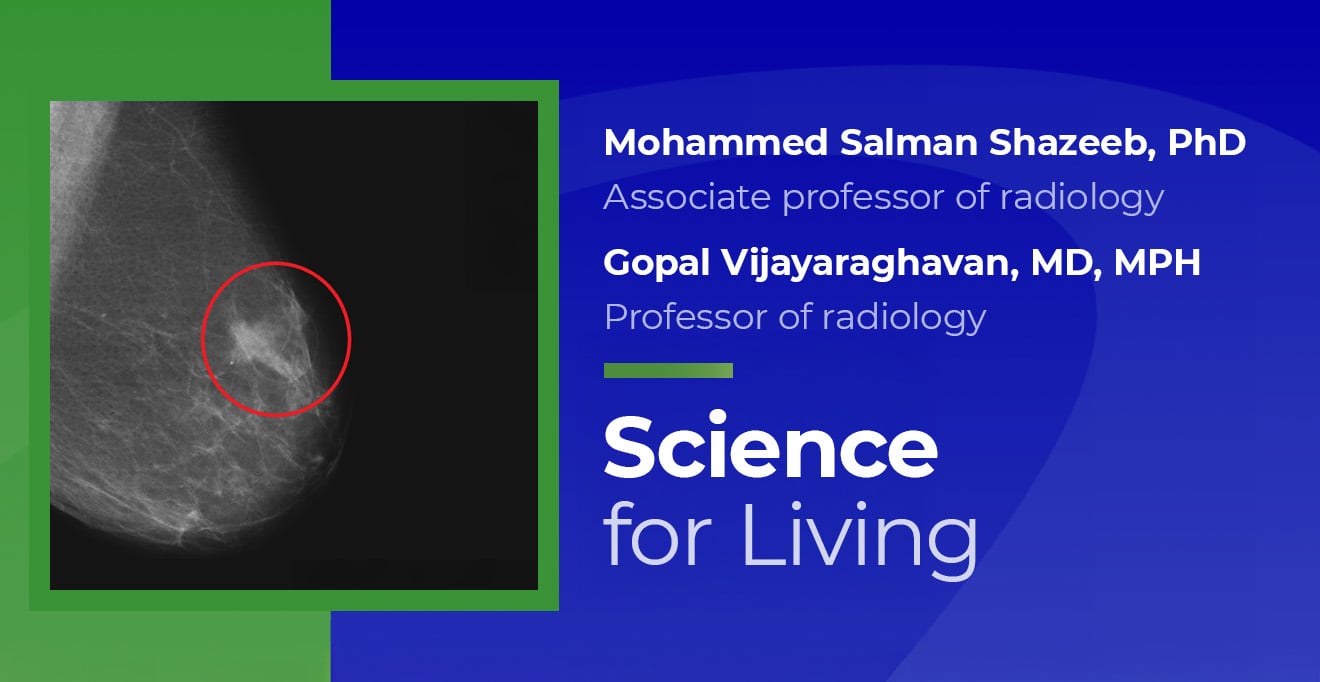
Dr. Mohammed Salman Shazeeb, an associate professor of radiology, alongside Dr. Gopal Vijayaraghavan, professor of radiology, is part of a team evaluating an AI-based risk assessment model. This model was developed in partnership with researchers from the Massachusetts Institute of Technology. Funded by grants from state agencies and the Breast Cancer Research Foundation, the tool analyzes routine screening mammograms and generates a risk score that estimates a woman’s likelihood of developing breast cancer over the next several years.
A Targeted Approach to Supplemental Screening
Rather than recommending additional imaging for all patients, the AI risk score enables researchers to identify a smaller group of women who are most likely to benefit from further testing.
“Among the approximately 6 to 7 percent of women who scored above our risk threshold, we invited them for contrast-enhanced breast MRI,” Dr. Shazeeb explained. “It’s remarkable that all had normal screening mammograms, yet MRI detected cancers in some that would have otherwise gone unnoticed.”
In the initial group of 145 study participants, MRI revealed four additional cancers that mammography missed. This detection rate is significantly higher than what standard mammography typically uncovers over a comparable number of women.
“The tool is trained for performance, not understanding. That’s why these tools are designed to augment, not replace, clinical judgment.”
“MRI continues to be the gold standard for identifying many breast cancers,” Dr. Vijayaraghavan stated. “However, it is costly, time-consuming, and not practical for annual use for everyone. This tool could help us direct resources toward women at the highest risk, making early detection more efficient and tailored.”
How AI “Sees” Risk
AI technology can identify imaging features that are too minute for the human eye, utilizing patterns learned from extensive datasets. This advanced pattern recognition is a key factor in revealing risk signals that may otherwise elude clinicians.
“The AI can analyze many more features in an image than a radiologist can visually discern,” Dr. Vijayaraghavan noted. “However, it does not process information the way a physician would. The tool is optimized for performance, not understanding. Thus, it is meant to complement, not replace, clinical judgment.”
Human oversight remains crucial to interpret the AI’s findings and to ensure that insignificant results are not inadvertently flagged. Continued research is essential to establish the right balance between sensitivity and specificity, and larger studies will be necessary to validate the tool across diverse populations.
Challenges and Future Steps
While initial results are encouraging, several challenges must be addressed before this technology can become a standard part of clinical practice. Issues such as obtaining FDA approval, formulating reimbursement policies, and ensuring equitable access to care all need careful consideration before integrating AI-guided risk assessment into routine screenings.
“Before widespread application can occur, we need larger-scale validation and data on real-world implementation,” Dr. Shazeeb emphasized. “It’s also critical to ensure its efficacy across different populations and varied mammography systems.”
Engaging patients is another vital aspect of this research. While reactions to AI’s role in imaging are varied, Sara Schiller, senior research program manager in the Department of Radiology, shared that many women are eager to contribute, particularly if they have family histories of breast cancer. “They want to help advance research and generally do not have reservations about AI itself,” she said.
A Personalized Future for Screening
Experts stress that while AI is not a cure-all, it presents a promising tool for customizing screening based on individual risk levels.
“The goal isn’t to replace mammograms,” Dr. Vijayaraghavan clarified. “It is to provide an additional layer of insights that can assist in detecting cancers earlier, when treatment is more effective and less invasive.”
As research progresses, tools like this could lead to a more personalized approach to breast cancer screening, integrating state-of-the-art technology with clinical expertise to enhance outcomes for women.