The recent advancements in technology are reshaping our understanding of human anatomy, particularly in the realm of forensic science and bioarchaeology. Researchers have uncovered new details about the complexities of human pelvic anatomy, emphasizing the adult iliac auricular surface. This study, published in the International Journal of Legal Medicine, combines geometric morphometrics and machine learning to explore the subtle asymmetries and sexual dimorphisms in this significant anatomical feature. The findings are poised to enhance the analysis of human remains, offering greater accuracy and context in various scientific fields.
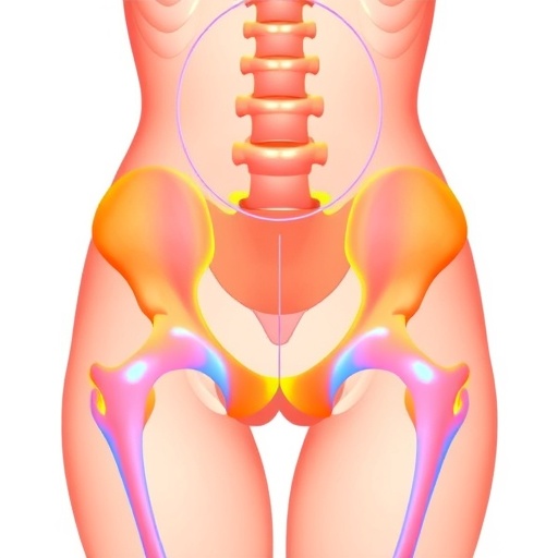
The iliac auricular surface, an essential part of the pelvic girdle, serves as a mechanical junction between the ilium and sacrum, facilitating load transfer from the spine to the lower limbs. While variations such as shape asymmetries and sexual dimorphisms have been acknowledged, quantifying these features has proven challenging due to their subtlety and individual variability. Traditional morphometric techniques often depend on manual landmarking and subjective judgments, which can introduce biases and hinder reproducibility. The application of computational geometric morphometrics represents a significant shift in this landscape.
Geometric morphometrics provides a detailed and quantitative framework for describing shape by capturing landmark configurations and analyzing their spatial arrangements. By utilizing high-resolution 3D models of the iliac auricular surface, the research team generated exact shape descriptors that illustrate individual variations in remarkable detail. This advanced method allowed them to identify consistent patterns of asymmetry and differentiate these from sexual dimorphic characteristics using strong statistical support, addressing challenges that previously complicated interpretation.
Machine learning algorithms further augmented the analytical framework, streamlining pattern recognition and classification tasks that would be overly complex for manual methods. The study utilized supervised learning techniques to develop models based on labeled datasets representing male and female specimens with established anatomical data. The algorithms exhibited remarkable precision in categorizing sexes based solely on the morphological parameters extracted from the iliac auricular surfaces. This automated approach enhances both the speed of analysis and the consistency of results, laying the groundwork for standardized protocols in forensic and anthropological applications globally.
The significant findings align with biological realities and evolutionary history. The asymmetry observed in the auricular surfaces reflects the concept of fluctuating asymmetry, a phenomenon often associated with developmental stability and environmental pressures during growth. The research reveals that these subtle asymmetries hold measurable importance and differ between sexes, offering deeper insights into pelvic biomechanics and reproductive biology. Understanding how pelvic shape impacts locomotion and load-bearing capabilities has critical implications in both medical and evolutionary contexts.
Sexual dimorphism in the pelvis is well-documented, arising primarily from the evolutionary demands of childbirth and upright walking. This study’s sophisticated morphometric methods elucidate the specific morphological distinctions of the iliac auricular surfaces, fostering a deeper understanding of how male and female pelvises differ not only in general but also in localized shape traits. By accurately quantifying these differences, the research enhances sex estimation techniques, which are crucial for forensic identification, archaeological reconstructions, and clinical assessments.
Importantly, the machine learning models were validated with diverse datasets, incorporating specimens from numerous populations, which addresses concerns about anthropological bias and broadens the method’s applicability. This cross-population validation bolsters the utility of the findings, positioning this approach as a universally adaptable tool. By minimizing the subjective judgment inherent in conventional morphological classifications, the research paves the way for more equitable and scientifically grounded methods in both bioarchaeology and forensic anthropology.
The integration of geometric morphometrics and machine learning also opens new avenues in personalized medicine. A detailed understanding of pelvic morphology can inform clinical treatments, surgical procedures, and rehabilitation strategies tailored to individual anatomical variations. Additionally, it enhances biomechanical modeling for prosthetics development, sports science, and injury prevention, showcasing the interdisciplinary potential of this technological synergy.
On a theoretical level, these methodologies offer evolutionary biology the means to better trace phenotypic changes over time and across populations. By rigorously quantifying shape variations and asymmetries, researchers can explore how environmental factors, genetics, and lifestyle changes influence pelvic morphology. This can lead to a richer narrative of human evolutionary adaptation, linking anatomical modifications to functional and behavioral outcomes.
This study highlights the transformative potential of artificial intelligence in managing complex biological data. By using machine learning not simply as a tool, but as an integrated partner in discovery, the researchers illustrate a shift in scientific methodology—one that values data-driven insights alongside domain expertise. This model denotes a paradigm shift, enhancing reproducibility and establishing a new benchmark for future morphological research.
While the research showcases remarkable technological progress, it also underscores existing challenges. The precise acquisition of 3D imaging, criteria for landmark selection, and computational resource needs all require careful optimization and standardization for routine application. Future studies must address these elements to create efficient protocols accessible to forensic practitioners and researchers.
Moreover, the societal implications of improving sex estimation techniques are significant. Ensuring the ethical use of these technologies in legal and anthropological contexts requires thoughtful consideration of privacy, consent, and cultural sensitivities. Transparent methodologies and open scientific discourse will be essential for navigating these responsibilities.
In summary, the collaborative effort by Amendola, Navega, Barucci, and their colleagues represents a landmark achievement at the intersection of technology and anthropology, challenging the limits of our understanding of human skeletal variation. By employing geometric morphometrics and machine learning to decode the intricate patterns of asymmetry and sexual dimorphism present in the iliac auricular surface, this research offers a promising vision for the future of forensic science, bioarchaeology, and evolutionary biology. The enhancements in both scientific knowledge and practical applications have the potential to impact identity, medicine, and history significantly.
As these methodologies continue to evolve and spread, the opportunity for broader anthropological insights expands, providing a more profound understanding of human biology through a precise lens of shape, symmetry, and detailed analysis. This innovative approach marks a significant advancement in human skeletal analysis, combining computational prowess with anthropological expertise to unlock the stories that our bones tell.
Subject of Research: Examination of asymmetry and sexual dimorphism in the adult iliac auricular surface utilizing geometric morphometrics and machine learning.
Article Title: Tracing asymmetry and sexual dimorphism in the adult iliac auricular surface: a geometric morphometrics and machine learning approach.
Article References:
Amendola, M., Navega, D., Barucci, A. et al. Tracing asymmetry and sexual dimorphism in the adult iliac auricular surface: a geometric morphometrics and machine learning approach. Int J Legal Med (2026). https://doi.org/10.1007/s00414-026-03726-z
Image Credits: AI Generated
DOI: https://doi.org/10.1007/s00414-026-03726-z
Tags: advancements in bioarchaeology methods, AI in pelvic anatomy research, anthropological insights from AI, challenges in traditional morphometrics, forensic applications of pelvic analysis, geometric morphometrics in anthropology, iliac auricular surface analysis, innovative technologies in anatomy studies, machine learning in forensic science, precision in human remains analysis, quantifying pelvic shape variations, sexual dimorphisms in human anatomy